


Molecular and Cellular Immunology Core
Home
As the sole resource for flow cytometry, cell sorting and state-of-the-art immunological services in the western-most IDeA state, the Molecular and Cellular Immunology Core, through COBRE support, has accelerated research productivity, in terms of publications and extramural funding. Emphasis has also been applied to developing new or customized immunological methods for COBRE Investigators and other Core users. In addition, regularly scheduled training sessions are held to enrich the educational and mentoring experience for COBRE Investigators and other faculty and students across the university and broader research community. By centralizing immunological services and resources to this Core, COBRE Investigators and other researchers will be more efficiently served and the use of expensive equipment will be maximized.
Objective
Enhance the infrastructure for the Molecular and Cellular Immunology Core, in support of hypothesis-driven research projects that seek to gain new knowledge about emerging infectious diseases.
Specific Aim 1
- Enhance and streamline core operations.
Specific Aim 2
- Grow and diversify user base, capability, capacity and reach.
Specific Aim 3
- Strengthen core infrastructure.
George S.N. Hui, Ph.D.
Director, Molecular and Cellular Immunology Core
Professor
Department of Tropical Medicine, Medical Microbiology and Pharmacology
John A. Burns School of Medicine
Email: ghui [at] hawaii.edu
Alexandra Gurary, Ph.D.
Associate Director, Molecular and Cellular Immunology Core
Email: gurary [at] hawaii.edu
Services/Assays
Core Services:
- Flow Cytometry: Analysis and Sorting
- Cell Counting, Size, and Viability
- Multiplex Bead Assays: Luminex and CBA Platforms for DNA, Protein, and Antibodies Assays
- Immunospot Plate Reading: ELISpot and FRNT
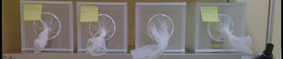
- Cell Irradiation
Flow Cytometry Assays:
- Cell Phenotyping
- Cell Cycle with Live or Fixed Cells
- Apoptosis
- FRET
- Calcium Flux
- Phosphorylation
- Intracellular Cytokine and Protein Detection
- Fluorescent Protein Expression
- Cell Population Isolation
- Single Cell Sorting
- Dye Dilution Assays
- Proliferation
Training opportunities
Experienced staff will provide hands-on training to investigators in conducting IBC/IACUC/CHS approved MCI research protocols. Please refer to MCI training document to help you get started on the process for obtaining clearance to work in the JABSOM Molecular and Cellular Immunology Core Facility.
Please contact Alexandra Gurary with any questions regarding flow cytometry, new applications or our facility at gurary[at]hawaii.edu, or you can give us a call at 808-692-1794 to discuss how the MCI core can complement your research.
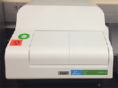
Equipment
 |
 |
 |
 |
 |
 |
Protocols/Procedures
As required by the COBRE EAC, we require that users submit a Service Request Form once a year for each research project, and a Sample Description Form for every appointment made with flow core operator, Alex Gurary.
Please fill out the service request and sample description form and email it to the Molecular and Cellular Immunology Core Facility.
All forms must be opened and completed with Adobe Reader.
Publications
Citations in Publications
Use of this core facility should be acknowledged in publications, abstracts, posters and oral presentations. The suggested verbiage is:
“Some of the services for this research were provided by the Molecular and Cellular Immunology Core, which is supported in part by grant P30GM114737 from the Centers of Biomedical Research Excellence (COBRE) program of the National Institute of General Medical Sciences, a component of the National Institutes of Health.”
2011-2012
- Darrow AL, Shohet RV, Maresh JG. Transcriptional analysis of the endothelial response to diabetes reveals a role for galectin-3. Physiological Genomics 2011 Oct 20;43(20):1144-52. [PMCID: PMC3217326, PMID: 21791638]
- Kelley JF, Kaufusi PH and Nerurkar VR. Dengue hemorrhagic fever-associated immunomediators induced via maturation of dengue virus nonstructural 4B protein in monocytes modulate endothelial cell adhesion molecules and human microvascular endothelial cells permeability. Virology 2012 Jan 20;422(2):326-37. [PMCID: PMC3273497, PMID: 22129847]
- Kelley JF, Kaufusi PH, Volper EM and Nerurkar VR. Maturation of dengue virus nonstructural protein 4B in monocytes enhances production of dengue hemorrhagic fever-associated chemokines and cytokines. Virology 2011 Sep 15;418(1):27-39. [PMCID: PMC3184475, PMID: 21810535]
- Ndhlovu LC, Lopez-Verges S, Barbour JD, Jones RB, Jha AR, Long BR, Schoeffler EC, Fujita T, Nixon DF, Lanier LL. Tim-3 marks human natural killer cell maturation and suppresses cell-mediated cytotoxicity. Blood 2012 Mar 1. [PMID: 22383801]
Ohta K, Yamamoto M, Lin Y, Hogg N, Akiyama H, Behringer RR, Yamazaki Y. Male Differentiation of Germ Cells Induced by Embryonic-Age-Specific Sertoli Cells in Mice. Biology of Reproduction 2012 Jan 18. [PMID: 22262692]
Pusic K, Xu H, Stridiron A, Aguilar Z, Wang A and Hui G. Blood stage merozoite surface protein conjugated to nanoparticles induce potent parasite inhibitory antibodies. Vaccine 2011 Nov 8;29(48):8898-908. [PMCID: PMC3202662, PMID: 21963870]
- Pusic KM, Hashimoto CN, Lehrer A, Aniya C, Clements DE and Hui GS. T cell epitope regions of the P. falciparum MSP1-33 critically influence immune responses and in vitro efficacy of MSP1-42 vaccines. PLoS One 2011;6(9):e24782. [PMCID: PMC3172285, PMID: 21931852]
- Sumoza-Toledo A, Lange I, Cortado H, Bhagat H, Mori Y, Fleig A, Penner R, Partida-Sanchez S. Dendritic cell maturation and chemotaxis is regulated by TRPM2-mediated lysosomal Ca2+ release. FASEB journal 2011 Oct;25(10):3529-42. [PMCID: PMC3177582, PMID: 21753080]
2010-2011
- de Alwis R, Beltramello M, Messer WB, Sukupolvi-Petty S, Wahala WM, Kraus A, Olivarez NP, Pham Q, Brian J, Tsai WY, Wang WK, Halstead S, Kliks S, Diamond MS, Baric R, Lanzavecchia A, Sallusto F and de Silva AM. In-depth analysis of the antibody response of individuals exposed to primary dengue virus infection. PLoS neglected tropical diseases 2011 Jun;5(6):e1188. [PMCID: PMC3119640, PMID: 21713020]
- Chen I, Kaufusi P, and Erdem G. Emergence of Erythromycin and Clindamycin-resistant Streptococcus pyogenes emm 90 strains in Hawaii. Journal of Clinical Microbiology. 2011 Jan;49(1):439-41. [PMCID: PMC3020461, PMID: 21068284]
Garcia AF, Abe LM, Erdem G, Cortez CL, Kurahara D, and Yamaga K. An insert in the covS gene distinguishes a pharyngeal and a blood isolate of Streptococcus pyogenes found in the same individual. Microbiology. 2010 Oct;156(Pt 10):3085-95. [PMCID: PMC3068697, PMID: 20634239]
Kumar M, Verma S, Nerurkar VR. Pro-inflammatory cytokines derived from West Nile virus (WNV)-infected SK-N-SH cells mediate neuroinflammatory markers and neuronal death. Journal of Neuroinflammation. 2010 Oct 31;7:73. [PMCID: PMC2984415, PMID: 21034511]
Lin SR, Zou G, Hsieh SC, Qing M, Tsai WY, Shi PY, Wang WK. The stem region of envelope protein of dengue virus is involved in virus assembly and entry. Journal of Virology. 2011 May;85(10):5159-71. [PMCID: PMC3126166, PMID: 21367896]
Verma S, Hoffmann FW, Kumar M, Huang Z, Roe K, Nguyen-Wu E, Hashimoto AS, Hoffmann PR. Selenoprotein K knockout mice exhibit deficient calcium flux in immune cells and impaired immune responses. Journal of Immunology. 2011 Feb 15;186(4):2127-37. [PMCID: PMC3088479, PMID: 21220695]
Verma S, Kumar M, Nerurkar VR. Cyclooxygenase-2 inhibitor blocks the production of West Nile virus-induced neuroinflammatory markers in astrocytes. Journal of General Virology. 2011 Mar;92(Pt 3):507-15. [PMCID: PMC3081232, PMID: 21106803]
Zhang L, Strianese O, Gaudino G, Morris P, Pass HI, Nerurkar VR, Bocchetta M, Carbone M. Tissue tropism of SV40 transformation of human cells: role of the viral regulatory region and of cellular Notch-1. Genes and Cancer. 2010 Oct;1(10):1008-20. [PMCID: PMC3092263, PMID: 21779427]
2009-2010
Imrie A, Roche C, Zhao Z, Bennett S, Laille M, Effler P, Cao-Lormeau VM. Homology of complete genome sequences for dengue virus type-1, from dengue-fever- and dengue-haemorrhagic-fever-associated epidemics in Hawaii and French Polynesia. Annals Tropical Medicine and Parasitology. 2010 Apr;104(3):225-35. [PMCID: PMC3084289, PMID: 20507696]
Lieberman MM, Nerurkar VR, Luo H, Cropp B, Carrion R, Jr., de la Garza M, Coller BA, Clements D, Ogata S, Wong T, Martyak T, Weeks-Levy C. Immunogenicity and protective efficacy of a recombinant subunit West Nile virus vaccine in rhesus monkeys. Clinical and Vaccine Immunology: CVI 2009 Sep;16(9):1332-7. [PMCID: PMC2745014, PMID: 19641099]
Rai MA, Nerurkar VR, Khoja S, Khan S, Yanagihara R, Rehman A, Kazmi, SU, Syed HA. Evidence for a founder effect among HIV-1-infected injection drug users (IDUs) in Pakistan. BMC Infectious Diseases. 2010 Jan 12;10:7. [PMCID: PMC2820481, PMID: 20064274]
Verma S, Kumar M, Gurjav U, Lum S, Nerurkar VR. Reversal of West Nile virus-induced blood-brain barrier disruption and tight junction proteins degradation by matrix metalloproteinases inhibitor. Virology. 2010 Feb 5;397(1):130-8. [PMCID: PMC3102050, PMID: 19922973]
Flow Cytometry Basics
GUIDE TO FLOW CYTOMETRY AND ASSAYS
The acronym FACS (Fluorescence Activated Cell Sorting) and flow cytometry are used interchangeably. FACS is a powerful method used to study and purify cells. FACS has a wide application in immunology and cell biology and other fields of biology.
Individual cells held in a thin stream of fluid are passed through one or more laser beams cause light to scatter and fluorescent dyes to emit light at various frequencies. Photomultiplier tubes (PMT) convert light to electrical signals and cell data is collected. Cell sub-populations are identified and sorted at high purity (~100%). FACS instruments generate three types of data:
- Forward Scatter (FSc) - Approximate cell size
- Fluorescent Labeling - Used to investigate cell structure and function
- Side or Orthogonal Scatter (SSc) - Cell complexity or granularity
Scatter
Forward and side scatter are used for preliminary identification of cells. In a peripheral blood sample, lymphocyte, monocyte and granulocyte populations can be defined on the basis of forward and side scatter. Forward and side scatter are used to exclude debris and dead cells.
Fluorescence
Fluorescent labeling allows investigation of cell structure and function. Cell auto fluorescence is generated by labeling cell structures with fluorescent dyes. FACS collect fluorescence signals in one to several channels corresponding to different laser excitation and fluorescence emission wavelength.
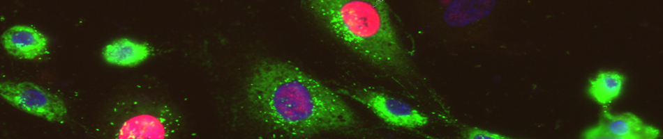
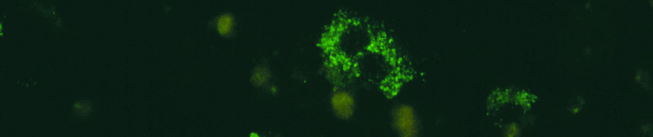
Immunofluorescence, the most widely used application, involves the staining of cells with antibodies conjugated to fluorescent dyes such as fluorescein and phycoerythrin. This method is often used to label molecules on the cell surface, but antibodies can be directed at targets in cytoplasm.
In direct immunofluorescence an antibody to a molecule is directly conjugated to a fluorescent dye (such as lymphocyte surface marker CD4). Cells are stained in one step.
In indirect immunofluorescence the primary antibody is not labeled. A second fluorescently conjugated antibody is added which is specific for the first antibody. For example, if the anti-CD4 antibody was a mouse IgG then the second antibody could beat rat antibody raised against mouse IgG. Immunofluorescence examples:
- Quantifying CD4 and CD8 subsets of T lymphocytes
- Intracellular cytokine staining
The use of Flow Cytometry can be divided into two broad categories, analysis and cell sorting.
Analysis
The ability of flow cytometers to evaluate cells at an extremely rapid rate (e.g. up to 20,000 events per second) makes this technology ideally suited for the reliable and accurate quantitative analysis of selected physical properties of cells of interest. The sensitivity of these instruments for detecting the presence of molecules expressed at low levels is impressive; given high quality cell preparations and reagents, as few as 50 molecules per cell may be detected.
Cell Sorting
One of the properties of the larger flow cytometers is the ability to electronically deflect cells with preset, defined properties into a separate collection tube. For cell purification, flow cytometry is especially well suited for applications requiring high purity. Because multiple fluorochromes (e.g. up to eight distinct fluorescent probes reacting with different cell associated molecules) can be assessed simultaneously, cell sorting by flow cytometry can separate complex mixtures of cells on the basis of multiple marker expression.
DNA Staining
FACS is used to study DNA cell content. Propidium iodide (PI) and Hoechst dyes bind to DNA and become fluorescent. PI cannot enter live cells and is included in immunofluorescent staining protocols to identify dead cells. Some Hoechst dyes can enter live cells. DNA staining can be used to study the cell division cycle. Relative DNA content shows the proportion of cells in G1, G2 and S phases. Apoptotic cells show characteristic smear on DNA staining. DNA staining examples:
- Studying changes in cell cycle in mutant cell
- Measuring apoptosis in cells after irradiation
Calcium Flux and Other Metabolic Studies
FACS can be used to investigate cell biology. Calcium flux can be measured using Indo-1 markers. This can be combined with immunofluorescent stain. For example, identify T cell subpopulations by immunofluorescence and measure calcium flux in response to an activating signal. Rhodamine-123 stains mitochondrial membranes is used to measure cellular activation. Rhodamine-123 is rapidly pumped out of some cells (for example hematopoietic stem cells).
CFSE binds to cell membranes and equally distributes when cells divide. Cell divisions in a period of time can then be counted. For example, labeling a population of cells with CFSE in vitro and reintroduce them in vivo. After a few days, the cells could be sampled and the amount of division measured. For example: Metabolic characteristics such as calcium flux, mitochondrial activity, pH, and free radical production can be measured in live cell populations in real time.
Gene Expression and Transfection
FACS is used to measure gene expression in cells transfected with recombinant DNA. This is achieved directly by labeling the protein product, or indirectly by using a reporter gene in the construct. Direct immunofluorescent labeling allows quantification of the product, and is suitable for relatively abundant proteins expressed on the cell surface. Indirect detection by reporter genes allows detection of transfectants at lower levels which cannot be detected easily by immunofluorescence. Examples of reporter genes are beta galactosidase and Green Fluorescent Protein (GFP). Beta galactosidase activity can be detected by FACS using fluorogenic substrates such as fluorescein digalactoside (FDG). FDG is introduced into cells by hypotonic shock and is cleaved by the enzyme to generate a fluorescent product trapped within the cell. One enzyme can generate a large amount of fluorescent product.
Cells expressing GFP constructs will fluoresce without addition of a substrate. GPF mutants are available which have different excitation frequencies, but emit fluorescence in the same channel. In a two-laser FACS, it is possible to distinguish cells which are excited by different lasers and assay two transfections at the same time.
- Expression of proteins can be measured directly by FACS.
- Transfection efficiency can be accurately determined.
- Transfection assays can be combined with staining and sorting for other markers.
- Transfected cells can be purified for analysis or use.
Dyes
Fluorescent dyes allow analyses of simultaneous parameters that can refine cell subpopulations. The fluorescent dyes that you can use will depend upon instrument used.
When different fluorescent dyes are used, signal spillover can occur between fluorescence channels. This needs to be corrected by setting compensation.
Setting correct compensation is important for obtaining accurate results when using multiple colors. In our facility we recommend use of compensation beads, when possible. Please contact Alexandra Gurary at gurary[at]hawaii.edu for further information.
Links
Contact Us
Please contact George Hui or Alexandra Gurary with any questions regarding flow cytometry and new applications or questions about the Molecular and Cellular Immunology Core.
Director:
George Hui
Email: ghui[at]hawaii.edu
Core Supervisor:
Alexandra Gurary
Phone: 808-692-1794
Email: gurary[at]hawaii.edu