


Publication Detail
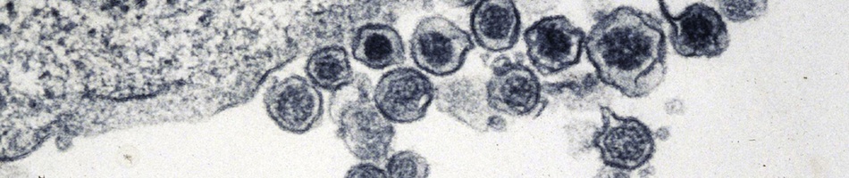
Cryo-electron tomography of rubella virus.
Battisti AJ, Yoder JD, Plevka P, Winkler DC, Prasad VM, Kuhn RJ, Frey TK, Steven AC, Rossmann MG.
Citation
Battisti AJ, Yoder JD, Plevka P, Winkler DC, Prasad VM, Kuhn RJ, Frey TK, Steven AC, Rossmann MG. (2012) Cryo-electron tomography of rubella virus. Journal of Virology 86(20):11078-11085.
Abstract
Rubella virus is the only member of the Rubivirus genus within the Togaviridae family and is the causative agent of the childhood disease known as rubella or German measles. Here, we report the use of cryo-electron tomography to examine the three-dimensional structure of rubella virions and compare their structure to that of Ross River virus, a togavirus belonging the genus Alphavirus. The ectodomains of the rubella virus glycoproteins, E1 and E2, are shown to be organized into extended rows of density, separated by 9 nm on the viral surface. We also show that the rubella virus nucleocapsid structure often forms a roughly spherical shell which lacks high density at its center. While many rubella virions are approximately spherical and have dimensions similar to that of the icosahedral Ross River virus, the present results indicate that rubella exhibits a large degree of pleomorphy. In addition, we used rotation function calculations and other analyses to show that approximately spherical rubella virions lack the icosahedral organization which characterizes Ross River and other alphaviruses. The present results indicate that the assembly mechanism of rubella virus, which has previously been shown to differ from that of the alphavirus assembly pathway, leads to an organization of the rubella virus structural proteins that is different from that of alphaviruses.
| Link: | http://www.ncbi.nlm.nih.gov/pubmed/22855483 |
| PMID: | 22855483 |
| PMCID: | PMC3457135 |