


Publication Detail
Clinical and imaging findings in an infant with Zika embryopathy.
Culjat M, Darling SE, Nerurkar VR, Ching N, Kumar M, Min SK, Wong R, Grant L, Melish ME.
Citation
Culjat M, Darling SE, Nerurkar VR, Ching N, Kumar M, Min SK, Wong R, Grant L, Melish ME. (2016) Clinical and imaging findings in an infant with Zika embryopathy. Clinical Infectious Diseases
Abstract
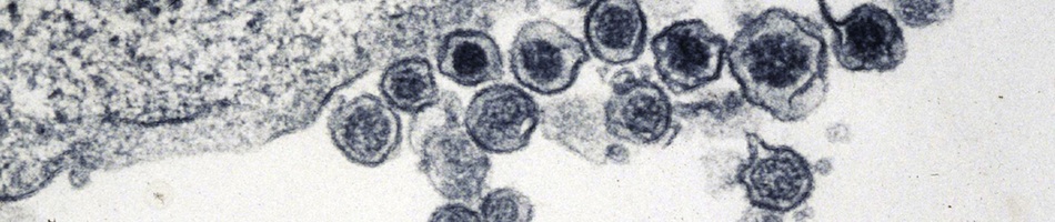
Recent Zika virus (ZIKV) outbreaks have been associated with an increased incidence of neonatal microcephaly. Subsequently, tropism for the brain was established in human fetal brain tissue. We present the first congenital ZIKV infection in the United States, confirmed by high ZIKV IgM antibody titers in serum and cerebrospinal fluid. The phenotypic characteristics of the patient fall within fetal brain disruption sequence, suggesting impaired brain development in the second half of gestation. Brain imaging revealed an almost agyric brain with diffuse parenchymal calcifications, hydrocephalus ex vacuo, and cerebellar hypoplasia. Ophthalmologic examination revealed macular pigment stippling and optic nerve atrophy. Liver, lungs, heart and bone marrow were not affected. The patient had progressive neurologic deterioration in the first month of life. The discovery of ZIKV infection in human fetal brain tissue along with serologic confirmation proves the vertical transmission of ZIKV. Therefore, ZIKV has joined the group of congenital infections.
| Link: | http://www.ncbi.nlm.nih.gov/pubmed/27193747 |
| PMID: | 27193747 |
| PMCID: |